Sketch And Label Of A Cross Section Of A Long Bone / Sketch And Label Of A Cross Section Of A Long Bone Cross Section Of Right Kidney Photograph By Science Source : Continue to label this drawing as you explore the inside of the bone.
byAdmin•
0
Sketch And Label Of A Cross Section Of A Long Bone / Sketch And Label Of A Cross Section Of A Long Bone Cross Section Of Right Kidney Photograph By Science Source : Continue to label this drawing as you explore the inside of the bone.. Create a drawing of the bone section in your laboratory journal and label the areas listed above. 1) from a mechanical standpoint, bone is historically the most studied tissue, and 2) due to 1) and the simpler behavior of bone compared to soft tissues, more is known about bone mechanics in relation to its structure. Plates of cartilage, also known as growth plates which allow the long bones to grow during childhood. Draw and label the following structures as they appear using the 10x objective o bone marrow o bony trabeculae activity 5.2.3: Two types of bone tissues in cross section of a long bone :
This photo shows a cross section through bone. Lamellar bone makes up the compact or cortical bone in the skeleton, such as the long bones of the legs and arms. Draw and label a longitudinal section of a long bone. Growth in length of a bone occurs at the 4. The digital cushion sits just behind the pedal bone and above the sensitive frog.
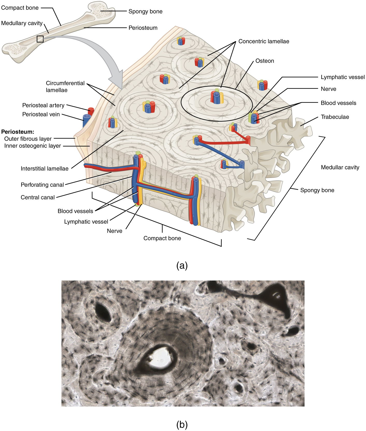
Skeletal Muscle Cross Section from medcell.med.yale.edu The periosteum contains many strong collagen fibers that are used to firmly anchor tendons and muscles to the bone for movement. Related posts of cross section of a long bone bone test anatomy and physiology. Smartdraw includes 1000s of professional healthcare and anatomy chart templates that you can modify and make your own. A long bone is a bone that has greater length than width. Draw a cross section of compact/osteon bone labeling all microscopic structures. The central haversian canal, and horizontal canals (perforating/ volkmann's) canals contain blood vessels and nerves from the periosteum. 1) from a mechanical standpoint, bone is historically the most studied tissue, and 2) due to 1) and the simpler behavior of bone compared to soft tissues, more is known about bone mechanics in relation to its structure. Bone test anatomy and physiology 12 photos of the bone test anatomy and physiology anatomy and physiology bone lab test, anatomy and physiology bone markings test, anatomy and physiology bone practical test, anatomy and physiology bone tissue test, anatomy and physiology test on bone tissue, bone, anatomy and.
Sketch and label a cross section of a bone.
External circumferential lamellae, osteon, central canal, perforating canals, lacuna, canaliculi, concentric lamellae. Cross section of long bone. Use the internet or a reference textbook to help you identify the external features of long bone listed below. The diaphysis and the epiphysis. The diaphysis of a long bone is composed of bone tissue while the epiphysis is made of 3. Related posts of cross section of a long bone bone test anatomy and physiology. Draw a cross section of compact/osteon bone labeling all microscopic structures. In the space provided, draw a longitudinal section of a long bone and label the following parts: Bone test anatomy and physiology 12 photos of the bone test anatomy and physiology anatomy and physiology bone lab test, anatomy and physiology bone markings test, anatomy and physiology bone practical test, anatomy and physiology bone tissue test, anatomy and physiology test on bone tissue, bone, anatomy and. The osteocytes are arranged in concentric rings of bone matrix called lamellae (little plates), and their processes run in interconnecting canaliculi. 1) from a mechanical standpoint, bone is historically the most studied tissue, and 2) due to 1) and the simpler behavior of bone compared to soft tissues, more is known about bone mechanics in relation to its structure. Chapter 6 bones and skeletal tissues flashcards quizlet. Learners should accurately draw a long bone, resembling that in figure 6.24.
A long bone is a bone that has greater length than width. In the space provided, draw a longitudinal section of a long bone and label the following parts: Area between the diaphysis and epiphysis at both ends of the bone. The outside of a bone is covered in a thin layer of dense irregular connective tissue called the periosteum. Long bones can have multiple epiphyses that are found at the ends of bone.
Bone Structure Anatomy And Physiology from opentextbc.ca There is a printable worksheet available for download here so you can take the quiz with pen and paper. Once we stop growing (between 18. Continue to label this drawing as you explore the inside of the bone. Lamellar bone makes up the compact or cortical bone in the skeleton, such as the long bones of the legs and arms. Draw and label the following structures as they appear using the 10x objective o bone marrow o bony trabeculae activity 5.2.3: Chapter 6 bones and skeletal tissues flashcards quizlet. It is located between the elbow joint and the shoulder. We start our section on tissue structure function with bone tissue.
The diaphysis and the epiphysis.
Long bones have a thick outside layer of compact bone and an inner medullary cavity containing bone marrow. Cross section of long bone. Make a pencil sketch and use markers or colored pencils to add details. The humerus is the long bone in the upper arm. Cartilaginous area at the ends of long bones where lengthwise growth takes place in the immature skeleton. Describe how this is live tissue which is both strong and slightly flexible osteons are the structural unit of compact bone, and enable to the bone to be able to bare weight with the way they are structured. Learners should accurately draw a long bone, resembling that in figure 6.24. Area between the diaphysis and epiphysis at both ends of the bone. You need to get 100% to score the 10 points available. Then, fill in the table below to describe each. This photo shows a cross section through bone. The diaphysis and the epiphysis. Two types of bone tissues in cross section of a long bone :
Proximal epiphysis, distal epiphysis, diaphysis, metaphysis, medullary cavity, epiphyseal line 2. The central haversian canal, and horizontal canals (perforating/ volkmann's) canals contain blood vessels and nerves from the periosteum. Bone test anatomy and physiology 12 photos of the bone test anatomy and physiology anatomy and physiology bone lab test, anatomy and physiology bone markings test, anatomy and physiology bone practical test, anatomy and physiology bone tissue test, anatomy and physiology test on bone tissue, bone, anatomy and. Lamellar bone makes up the compact or cortical bone in the skeleton, such as the long bones of the legs and arms. Forms the larger rounded ends of long bones.
Sketch And Label Of A Cross Section Of A Long Bone Sketch And Label Of A Cross Section Of A Long Bone Long Bone Cross Section Worksheet Teaching Resources As The from i2.wp.com A long bone has a shaft and 2 ends. A long bone has two parts: The osteocytes are arranged in concentric rings of bone matrix called lamellae (little plates), and their processes run in interconnecting canaliculi. Label the haversian canal, osteocyte (mature bone cell) in lacuna, and canaliculi. Cartilaginous area at the ends of long bones where lengthwise growth takes place in the immature skeleton. Only the bottom portion of this bone extends as far as the hoof capsule. Use the internet or a reference textbook to help you identify the external features of long bone listed below. Proximal epiphysis, distal epiphysis, diaphysis, metaphysis, medullary cavity, epiphyseal line 2.
Label lines should not cross ;
Long bones can have multiple epiphyses that are found at the ends of bone. Cartilaginous area at the ends of long bones where lengthwise growth takes place in the immature skeleton. Shop the edit of floral dresses, dream jeans and fresh shoes now, and stay tuned for a lot more exciting topshop stuff to come. The diaphysis is the tubular shaft that runs between the proximal and distal ends of the bone. Proximal epiphysis, distal epiphysis, diaphysis, metaphysis, medullary cavity, epiphyseal line 2. Plates of cartilage, also known as growth plates which allow the long bones to grow during childhood. As the names suggest compact bone looks compact and the spongy bone looks like sponges. Create a drawing of the bone section in your laboratory journal and label the areas listed above. Also known as the middle phalanx, the short pastern bone sits on top of the articulating joint of the pedal bone and underneath the long pastern bone. There is a printable worksheet available for download here so you can take the quiz with pen and paper. A long bone has two parts: External circumferential lamellae, osteon, central canal, perforating canals, lacuna, canaliculi, concentric lamellae. Related posts of cross section of a long bone bone test anatomy and physiology.